Home | Surgeries | Luxating Patella (Dislocated Kneecap) Surgery in Dogs and Cats
Luxating Patella (Dislocated Kneecap) Surgery in Dogs and Cats
Luxating Patella (Dislocated Kneecap) Surgery in Dogs and Cats
What is a luxating patella? The patella, or "kneecap," is normally located in a groove on the end of the femur (thigh bone). The term luxating means "out of place" or "dislocated." Therefore, a luxating patella is a kneecap that moves out of its normal location and may cause discomfort, lameness, or joint damage if left untreated.
Luxating patella, or dislocated kneecap, is one of the most common orthopedic conditions seen in dogs—especially small breeds like Pomeranians, Yorkshire Terriers, Chihuahuas, and Toy Poodles. At Canton Animal Hospital, we diagnose and treat patellar luxation using advanced orthopedic techniques and a compassionate, pet-focused approach.
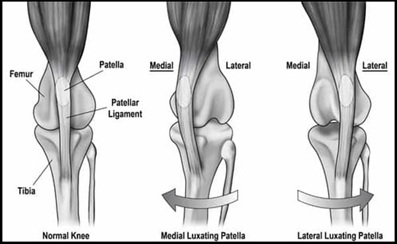
When a dog has patellar luxation, the kneecap (patella) slips out of its normal groove in the femur, leading to intermittent or chronic lameness, skipping gaits, or pain. Luxation can be medial (toward the inside) or lateral (toward the outside), and severity is graded from I to IV.
Causes & Risk Factors
Congenital malformation (most common cause)
Shallow femoral groove or misaligned limb anatomy
Traumatic injury to the stifle (knee joint)
Common in small and toy breeds, but also seen in large dogs
May occur in one or both knees
Featured Resources

We Welcome New Patients!
We're always happy to give your furry friend care at our hospital. Get in touch today!
Contact Us
Symptoms of Patellar Luxation
Dogs of any age or breed and either gender may be affected, but small- and toy-breed dogs are the most frequently diagnosed.
Medial patellar luxation is more common than lateral luxation, especially in small breeds.
Large-breed dogs may also be affected, although lateral luxation is more typical in these cases.
Toy breeds such as Maltese, Chihuahua, French Poodles, and Bichon Frise are genetically predisposed.
Abnormal positioning of the patellar ligament, particularly in bowlegged dogs, contributes to luxation.
Most affected dogs exhibit intermittent weight-bearing lameness or limping
Sudden yelp of pain during play or running
Hopping or skipping on one hind limb
Difficulty standing or climbing stairs
Bowlegged appearance or stiff-legged walking
Clicking or popping sounds from the knee
Grade IV luxation is associated with persistent lameness and obvious gait abnormalities.
Physical Exam Findings:
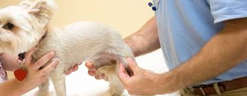
The diagnosis of medial patellar luxation is based on finding or eliciting medial patellar luxation during a physical examination. Physical findings vary and depend on the severity of luxation. Grades of Patellar Luxation:
Grade I
The patella can be luxated, but spontaneous luxation of the patella during normal joint motion rarely occurs. Manual luxation of the patella may be accomplished during physical examination, but the patella reduces when pressure is released. Flexion and extension of the joint are normal.
Grade II
Angular and torsional deformities of the femur may be present to a mild degree. The patella may be manually displaced with lateral pressure or may be luxate with flexion of the stifle joint. The patella remains luxated until the examiner reduces it or it is spontaneously reduced when the animal extends and de-rotates its tibia.
The proximal tibial tuberosity may be rotated up to 30 degrees with medial luxation and less so with lateral luxation. With the patella luxated medially, the hock is slightly abducted with the toes pointing medially (“pigeon toed”). With lateral luxation, the hock may be adducted with the toes pointing laterally (“seal-like”).
Grade III
The patella remains luxated medially most of the time but may be manually reduced with the stifle in extension. However, after manual reduction, flexion and extension of the stifle result in re-luxation of the patella. There is a medial displacement of the quadriceps muscle group. Abnormalities of the supporting soft tissue of the stifle joint and deformities of the femur and tibia may be present.
Torsion of the tibia and deviation of the tibial crest between 30 and 60 degrees from the cranial/caudal plane are present
Grade IV
There may be an 80- to 90-degree medial rotation of the proximal tibial plateau. The patella is permanently luxated and cannot be manually repositioned. The femoral trochlear groove is shallow or absent, and there is medial displacement of the quadriceps muscle group. Abnormalities of the supporting soft tissue of the stifle joint and deformities of the femur and tibia are notable.
Diagnosis
At Canton Animal Hospital, we perform a full orthopedic exam and gait evaluation to detect patellar instability. Radiographs (X-rays) are used to confirm the diagnosis and assess for associated joint or bone abnormalities.
Diagnostic Imaging:
With a grade III or grade IV luxation, standard craniocaudal and medial-to-lateral radiographs show the patella to be displaced medially,
Whereas with a grade I or grade II luxation the patella may be within the trochlear sulcus or may be displaced medially.
Full limb radiographs may show varus or valgus deformities and torsion of the tibia and femur.
Careful radiographic positioning is critical, as poor positioning results in false positive limb deformity on radiographs.
Laboratory Finding:
Consistent laboratory findings are not seen. Arthrocentesis demonstrates changes compatible with osteoarthritis.
Differential Diagnosis: Differential diagnoses include:
Avascular necrosis of the femoral head. (Legg-Calve-Perthes Disease)
Coxo-femoral luxation (Hip Joint Luxation),
Cranial cruciate ligament rupture.
Careful examination of the hip joint is essential because some patients with patellar luxation also have avascular necrosis of the femoral head or hip dysplasia.
Shortening of the limb because of hip luxation or femoral head osteotomy (FHO) will cause laxity of the quadriceps mechanism, enabling luxation of the patella in some cases. This usually resolves with treatment of the hip luxation and with time after FHO.
Treatment Options
Treatment depends on severity and impact on quality of life:
Conservative Management
Pain relief medications and joint supplements
Physical therapy and low-impact exercise
Weight management to reduce joint stress
Not typically recommended for Grades III–IV
Surgical Correction
Surgical Treatment: Numerous surgical techniques are aimed at restraining the patella within the trochlear groove.
Trochlear wedge or block recession
Tibial crest transposition
Medial fascial release
Lateral imbrication
Anti-rotational sutures,
Femoral osteotomy,
Tibial osteotomy,
Surgery is advised in symptomatic immature or young adult patients because intermittent patellar luxation may prematurely wear the articular cartilage of the patella.
Surgery is indicated at any age in patients showing lameness and is strongly advised in those with active growth plates because skeletal deformity may worsen rapidly.
The surgical techniques used in actively growing animals should not adversely affect skeletal growth.
Owners of dogs with bilateral grade IV patellar luxation should be warned of the likely need for multiple surgeries and probable continued lameness even after successful surgery because of the severity of the underlying long-bone abnormalities.
Our team ensures a customized surgical plan based on your pet’s anatomy, severity of luxation, and age.
Anatomical Consideration before Patella Surgery
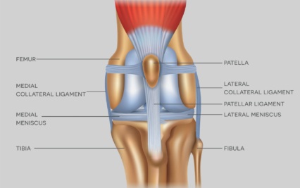
The extensor mechanism of the stifle joint is composed of the quadriceps muscle groups, patella, trochlear groove, straight patellar ligament, and tibial tuberosity.
The quadriceps muscle group is formed by the rectus femoris, vastus lateralis, vastus intermedius, and vastus medialis muscles.
The vastus medialis and vastus lateralis muscles are fixed to the patella by the medial and lateral parapatellar fibrocartilage. The fibrocartilage rides on the ridges of the femoral trochlea and, along with the medial and lateral retinacula, aids in patellar stability.
The medial and lateral retinacula are groups of collagen fibers that course from the fabella to blend with the medial and lateral parapatellar fibrocartilages, respectively.
The quadriceps muscle group extends the stifle joint and, along with the entire extensor mechanism, aids in stabilizing the stifle joint. The quadriceps muscle group converges as the patellar tendon on the patella and then continues distally as the straight patellar ligament.
The patella is a sesamoid bone embedded in the tendon of the quadriceps muscle. The inner articular surface is smooth and curved so as to fully articulate with the trochlea.
The normal gliding articulation of the patella and trochlea is necessary for maintaining the nutritional requirements of the trochlear and patellar articular surfaces.
The patella is also an essential component of the functional mechanism of the extensor apparatus. The patella maintains even tension when the stifle is extended and also acts as a fulcrum in a lever arm, increasing the mechanical advantage of the quadriceps muscle group.
The tibial tuberosity is located cranial and distal to the tibial condyles. Its location and prominence are important for the mechanical advantage of the extensor mechanism. The alignment of the quadriceps, patella, trochlea, patellar ligament, and tibial tuberosity must be normal for proper function. Malalignment of one or more of these structures may lead to patellar luxation.
Special anatomic considerations in patients with medial patellar luxation must be noted. When the patella is positioned medially, the patellar ligament must be identified before making a parapatellar incision to enter the joint.
The lateral capsule is stretched and thin, whereas the medial capsule is contracted and thickened. Thickening and contraction of the medial capsule are apparent if medial release is performed. The insertions of the caudal head of the sartorius muscle and the vastus medialis muscle should be identified. The medial ridge of the trochlear sulcus and the ventral surface of the patella may be worn.
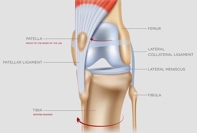
Surgical Considerations & Biomechanics
Luxating patella is a biomechanical disorder where the quadriceps mechanism is misaligned with the trochlear groove. To achieve lasting correction and patellar stability, multiple surgical techniques are often required.
Trochlear deepening procedures, such as wedge or block recession, are used to reshape and deepen the groove to better contain the patella.
Medial retinacular release may be performed to allow freer movement of the kneecap into its corrected alignment.
Tibial tuberosity transposition (TTT) realigns the patellar ligament with the center of the trochlear groove, correcting the directional pull of the quadriceps.
Lateral retinacular reinforcement techniques—such as suture tightening, fascia lata grafts, or capsular imbrication—help stabilize the patella post-reduction. However, these are not sufficient alone unless mechanical forces are properly corrected.
In severe cases with bony deformities (e.g., distal femoral varus or medial torsion of the tibia), a femoral osteotomy may be required to achieve correct alignment. This involves preoperative measurement and wedge osteotomy to realign the stifle and restore biomechanical balance.
Failure to neutralize abnormal joint forces can lead to stretch failure of soft tissue repairs and recurrence of luxation.
Patellar luxation also predisposes the stifle to other injuries, particularly cranial cruciate ligament (CCL) rupture. In small-breed dogs with medial patellar luxation (MPL), up to 41% also have concurrent CCL injuries. These dual pathologies increase surgical complexity and reinforce the need for early intervention and comprehensive repair.
Surgical Techniques:
Make a craniolateral skin incision 4 cm proximal to the patella and extend the incision 2 cm below the tibial tuberosity. Incise the subcutaneous tissue along the same line. Incise the lateral retinaculum and joint capsule to expose the joint.
Deepening of the Trochlear Groove- Trochleoplasty
Trochlear Chondroplasty
This “cartilage flap” technique is useful only in puppies up to 10 months of age. As an animal matures, the cartilage becomes thinner and more adherent to the subchondral bone, making flap dissection difficult.
A cartilage flap is elevated from the sulcus, the subchondral bone removed from beneath it, and the flap pressed back into the deepened sulcus. If the sulcus is not deep enough, the process is repeated. This results in a deepened trochlea, with maintenance of articular cartilage in the sulcus and with fibrocartilage or fibrous tissue at the incisional gaps. The cartilage flap survives. and experimental dogs have shown no adverse effects from the procedure.
Trochlear wedge Sulcoplasty
Trochlear wedge recession deepens the trochlear groove to restrain the patella and maintain the integrity of the patellofemoral articulation.
If necessary, remodel the free osteochondral wedge with rongeurs to allow the wedge to seat deeply into the new femoral groove.
Replace the free osteochondral wedge when the depth is sufficient to house 50% of the height of the patella.
The osteochondral wedge remains in place because of the net compressive force of the patella and friction between the cancellous surfaces of the two cut edges.
Trochlear block recession.
Trochlear block recession deepens the trochlear groove to restrain the patella and maintain the integrity of the patellofemoral articulation. Cut into the articular cartilage of the trochlea, making a block-shaped outline. Be sure that the width of the cut is sufficient to accommodate the width of the patella but preserves the trochlear ridges.
Deepen the recession in the trochlea by removing additional bone from the base of the groove. Replace the free osteochondral fragment when the depth is sufficient to house 50% of the height of the patella. The osteochondral wedge remains in place because of the net compressive force of the patella and friction between the cancellous surfaces of the two cut edges.
The medial joint capsule is thicker than normal and contracted in patients with a grade III or grade IV medial patellar luxation. In these patients, the medial joint capsule and retinaculum must be released to allow lateral placement of the patella.
Place a cruciate or simple interrupted suture that does not close the tissue gap when the patella is in the proper position. Placement of a loose suture will prevent iatrogenic lateral luxation.
Tibial tuberosity transposition
Make a lateral parapatellar incision through the fascia lata, and carry the incision distally onto the tibial tuberosity below the joint line.
Reflect the cranialis tibialis muscle from the lateral tibial tuberosity and tibial plateau to the level of the long digital extensor tendon.
Complete the osteotomy in a proximal to distal direction. Do not transect the distal periosteal attachment.
The degree of lateral movement of the tibial tuberosity is subjective, but it is based on the longitudinal realignment of the tuberosity relative to the trochlear groove.
Once the site of relocation is chosen, remove a thin layer of cortical bone with a rasp or osteotome. Lever the tibial tuberosity into position and stabilize it with one or two small Kirschner wires directed caudally and slightly proximally.
Engage the caudal cortex, but do not exit the pin from the tibia caudally; if the pin protrudes too far from the caudal cortex of the tibia, persistent lameness results.
Security of the tibial transposition is increased by use of a figure-eight wire. The addition of a figure-eight wire is strongly recommended when performing tibial crest transposition in large-breed dogs and should be considered in all but the smallest patients. In large-breed dogs a lag screw may be used in place of the Kirschner wires.
Lateral imbrication.
For suture imbrication, place a polyester suture through the femoral-fabellar ligament and lateral parapatellar fibrocartilage.
Next, place a series of imbrication sutures through the fibrous joint capsule and lateral edge of the patella tendon.
With the leg in slight flexion, tie the femoral-fabellar suture and imbrication sutures.
If the patella is out of position most of the time, the retinaculum opposite the side of the luxation will be stretched; with medial luxation, there is redundant lateral retinaculum.
Once the patella has been reduced, excise the excess retinaculum and joint capsule, allowing tight closure of the arthrotomy.
Expected outcome
With proper surgical intervention, the prognosis is excellent for most dogs. Early treatment helps prevent progression to arthritis or irreversible joint damage. Dogs with bilateral luxation can have both knees repaired either simultaneously or in stages, depending on severity and health status.
Recurrent luxation after surgery has been reported in up to 50% of joints evaluated. However, most are grade I luxation that do not affect clinical function.
Most stifle joints function well enough that lameness is not apparent during examination, nor do clients report clinical dysfunction.
Most patients with recurrent luxation show re-luxation only on physical examination when manual force is used to displace the patella.
Overall, the prognosis for patients undergoing surgical correction of a grade I to III patellar luxation is excellent for return to normal limb function.
DJD progresses despite treatment, but it is not as severe as that associated with chronic CCL rupture.
Prognosis for patients with grade IV patellar luxation is guarded. Many joints require multiple surgery, and some patellae cannot be reduced without major corrective osteotomies.
Postoperative Care & Assessment:
Activity should be restricted to specific physical rehabilitation exercises and leash walking for 6 to 8 weeks; then the animal should be gradually returned to normal activity over a 6-week period.
Use of an E-collar to prevent licking or chewing the incision
Pain management and anti-inflammatories
Short leash walks only; no running or jumping
Because many of the small breeds are “jumpers,” padded bandage support for 10 to 14 days may be useful in the active patient.
Radiographs should be obtained at 4 and 8 weeks to evaluate healing of a tibial crest transposition.
Most dogs return to normal activity within 2–3 months and enjoy significantly improved mobility and quality of life.
Schedule a Consultation
If your dog is limping or showing signs of kneecap dislocation, schedule an orthopedic evaluation at Canton Animal Hospital. Our experienced team provides advanced diagnostics and surgical treatment for luxating patella and other joint issues.
Call us or visit www.cantonvets.com to request an appointment.
Frequently Asked Questions (FAQs)
Luxating patella can raise many questions for dog owners. Below are answers to some of the most commonly asked questions about kneecap dislocation in dogs
Featured Resources

We Welcome New Patients!
We're always happy to give your furry friend care at our hospital. Get in touch today!
Contact Us